


Traumatic avulsion
Traumatic avulsion
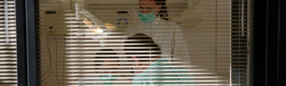
One Sunday morning a 13-year-old boy arrived at our office who, following a fall, had suffered the complete avulsion of the right central incisor, whose crown also showed a dentinal enamel fracture with exposure of the pulp (III class of Ellis), the dislocation of the left central incisor with dentinal enamel fracture without exposure of the pulp (Ellis class II) and the fracture of the right lateral. (Figure 1)

The avulsed element had been stored in milk but the time elapsed from the traumatic event was approximately 12 hours.
As per protocol, we placed the tooth in Hank's buffered saline solution and proceeded to perform the clinical examination and collect the anamnestic data.

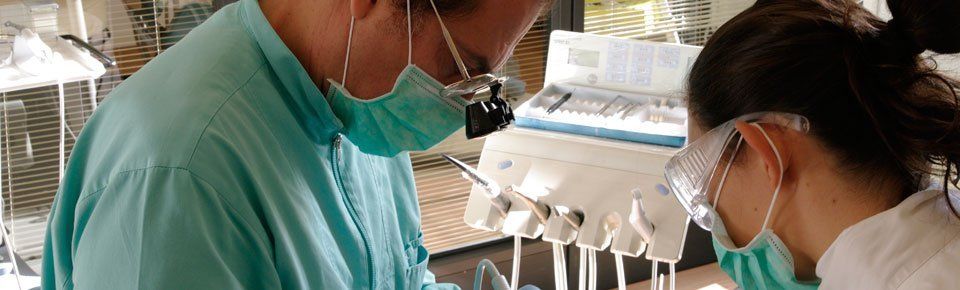
Since the avulsed tooth is a tooth with a fully formed apex and whose residence time outside the mouth was more than 60 minutes, we decided to carry out the root canal treatment outside the oral cavity, paying maximum attention, however, in canal irrigation. with disinfectants, not to damage the periodontal ligament by immersing the tooth every 2 minutes in buffered saline.

Before re-implantation, we performed the composite reconstruction of the other two fractured incisors.
We therefore dedicated ourselves to a delicate revision of the alveolus by lightly aspirating the clot that had formed.
The avulsed element was thus repositioned in the alveolus with a gradual compression until the dental alignment was reached and was thus kept in position for ten minutes.

The patient was then prescribed systemic antibiotics, chlorhexidine rinses and given strict oral hygiene instructions which are of paramount importance for as long as the splint is in place.

After 4 weeks of splint removal, the right incisor was perfectly stable (mobility grade 1) while the left incisor maintained its vitality.
Before the patient was discharged, the composite reconstructions were renewed to give a better aesthetic result. (figure 6)


LENCI DR. FEDERICO STUDIO DENTISTICO - Via PONTANO 7 - 80122 - Naples (NA)
Tel: +39 081 680650 | Fax: +39 081 680650 | E-mail: studiof.lenci@gmail.com
VAT Reg No 05748640637 | Legal Information
| Privacy and Cookie Policy
Designed by  |
Questa azienda è presente anche su
|
Questa azienda è presente anche su  e
e 
|
Questa azienda è presente anche su
e