Cone-Beam CT

Slide Title
Write your caption here
Button
Slide Title
Write your caption here
Button
Slide Title
Write your caption here
Button
CONE-BEAM CT
Accurate prevention strategies

In the medical field it is essential to carry out a correct diagnosis in order to set up a treatment plan that aims to have predictable and lasting results.
Dentists are already able to diagnose the main pathologies of the oral cavity with a simple visit: caries, periodontal disease, but when the diagnosis becomes less certain, for a more in-depth study, an analysis is necessary that allows us to study the tooth and the periodontium at 360 °
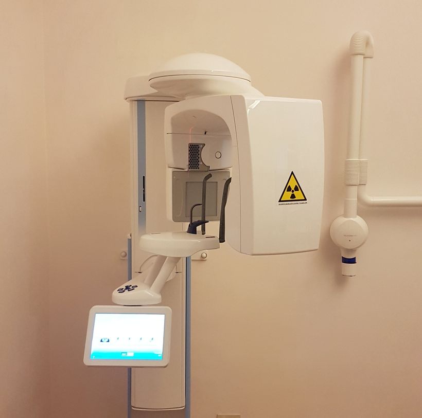
To meet this need, our practice has equipped itself with a carefully designed X-ray unit, Planmeca ProMax 3D Classic, which allows us to perform radiographic images in 2D (panoramic) or 3D (cone beam computed tomography: CBCT), with a radiation dose for the patient in accordance with the ALARA principle (lowest reasonably achievable level).
In addition, the pioneering Planmeca Ultra Low Dose protocol allows 3D images to be obtained, with an even lower dose, compared to standard 2D panoramic images, reducing the effective dose for the patient by up to 75-80%.
This new radiographic image processing protocol has changed our traditional procedures: in many cases a 2D (panoramic) examination is no longer justified, as the 3D ultra-low dose image allows for much more information with a similar dose of rays. .
In cases where the diagnosis should involve a narrower field of the oral cavity (only one tooth for endodontic causes), it will be possible to perform a selective radiographic examination for single dental or bone elements, this ROI reconstruction function (region of interest), allows a more precise diagnosis without the need for additional radiation for the patient.
Patients will be so happy to receive their diagnosis and treatment plan immediately, without the need to go to a specialized radiology center!
LENCI DR. FEDERICO STUDIO DENTISTICO - Via PONTANO 7 - 80122 - Naples (NA)
Tel: +39 081 680650 | Fax: +39 081 680650 | E-mail: studiof.lenci@gmail.com
VAT Reg No 05748640637 | Legal Information
| Privacy and Cookie Policy


